Flex-Axiom
Voici le seul résultat
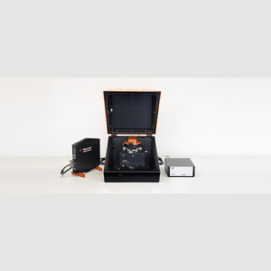
Nanosurf – Flex-Axiom — AFM for materials research
Versatility, performance and seamless application extensions
- Easy to use and high performance
- High resolution and low-noise design
- Modes and accessories to handle all your research needs
- Extremely modular to keep you productive
- Upgradable for life science and operation on large stages
Applications
- Imaging of samples in air and liquid
- Advanced mechanical, electrical and magnetical modes
- Electrochemical AFM
- Environmental control
- Scanning thermal microscopy and nano thermal analysis (SThM and Nano-TA)
- Advanced force spectroscopy
Lateral force microscopy on polystyrene-polybutadiene blend
The lateral force imaging mode was used to image local friction differences between the two polymers
Polystyrene and polybutadiene are shown to have quite different surface properties |
Overlay of difference between lateral deflection in forward and backward directions on top of topography. The difference is dominated by friction forces. Polystyrene (green areas) shows a higher friction than polybutadiene.System: FlexAFM with ES2 controller Scan size: 9 µm Cantilever: PPP-CONTSCROverlay of friction on topography |
 |
Overlay of average of lateral deflection in forward and backward directions on top of topography. The average is dominated by slope variations in the sample. The steepest slope (in red) is observed close to a large inset of polystyrene. The average shows no large color difference between the polymers in flatter areas.Overlay of slope on topography |
 |
Line section of lateral deflection difference and average of forward and backward scan directions. Line section of friction and slope signals |
Electrochemical AFM
This report demonstrates the capability of the FlexAFM in studies of charged solid-liquid interfaces
In order to carry out electrochemical(in-situ)AFM experiments, a conductive sample was mounted in an electrochemical liquid cell and connected to a lab-build bipotentiostat. We employed Clavilier-type Au(111) single crystal bead crystal electrodes with facets of micrometer-wide terraces. As examples, we studied the lifting of the Au(111)-(pxV3)reconstruction, surface oxidation as well as the growth and dissolution of copper clusters in sulfuric acid solution.
3D Copper cluster grown on Au(111)
KPFM using the Nanosurf FlexAFM
Kelvin probe force microscopy imaging
Kelvin Probe Force Microscopy (KPFM) is an extension of AFM. The technique was first published in 1991 by Nonnenmacher and coworkers. Using KPFM, images can be recorded that contain information on the local work function or local contact potential difference between tip and sample. Although all Nanite systems with an SPM S200 controller and all current Easyscan 2 AFM systems are in principle capable of performing KPFM, the FlexAFM has demonstrated best KPFM performance and is therefore the instrument of choice for this type of measurement. For a detailed description of requirements and procedures, please contact Nanosurf support. |
Topography. Scan Range: 10 µm x 10 µm. Z-range: 9 nm. |
 |
KPFM Signal. Scan range: 10 µm x 10 µm. Local charges that were placed on an insulating (oxide) surface layer in a "Swiss cross" pattern. Image courtesy: Marcin Kisiel, Thilo Glatzel and students of the Nanocurriculum of the University of Basel.KPFM signal (top) and simultaneously recorded Topography data (bottom) are shown. |