De retour à Microscopes

Nanosurf DriveAFM

Le DriveAFM, le nouvel instrument phare de Nanosurf utilise les dernières technologies pour offrir des  performances stables et haut de gamme. Il a été conçu pour répondre aux besoins d’une recherche de premier ordre, aujourd’hui et dans le futur.

  •  CleanDrive : excitation stable dans l’air et le liquide
  •  Bruit ultra-faible
  • Entraînement direct : imagerie haute résolution et grande zone de numérisation
  • Système entièrement motorisé : contrôle total via logiciel.
Cliquez ici pour télécharger plus de détails


Description

Performance without compromise

The DriveAFM, Nanosurf‘s new flagship instrument, utilizes the latest technology to deliver stable, high-end performance. It was designed to fulfill the needs of top notch research, today and in the future.
  • CleanDrive: stable excitation in air and liquid
  • Ultra-low noise
  • Direct drive: high-resolution imaging and large scan area
  • Fully motorized system: full control via software

CleanDrive: stability in air and liquid with photothermal excitation

Photothermal excitation of the cantilever provides unparalleled stability, a linear frequency response, and a high excitation bandwidth in air and liquid environments. These benefits allow measurements at multiple frequencies and highspeed applications and open new horizons for innovative new measurement modes (e.g. Cytomass Monitor).

These advantages are amplified in liquids since only the cantilever beam is excited and the liquid environment remains largely unperturbed. This results in clean resonance peaks and not the “forest of peaks” commonly seen with the piezo acoustic excitation of cantilevers. Furthermore, this method of exciting the cantilever is insensitive to changes in the environment and distance to the sample, making the whole measurement system much more stable.

The DriveAFM, with its small light spots, is compatible with the use of small cantilevers, which have several advantages that make them superior in performance. While they have the same spring constant as a conventional cantilever, small cantilevers show a significantly higher resonance frequency and operational bandwidth. Also, the noise performance is better. Due to the small dimensions, the sensitivity is increased, and hydrodynamic drag is decreased. All of this results in better imaging performance.

Ultra-low noise

The DriveAFM has a very low overall noise floor, which is achieved through a combination of a low-noise/low-coherence superluminescent diode and a low-noise/high-bandwidth photodetector used in the beam deflection detection module and the low-noise/high-bandwidth CX Controller. This is the basis for the stable, sensitive, and high-resolution imaging and force spectroscopy capability of the DriveAFM.


Ultra-low noise
Direct drive scanner

The DriveAFM exploits the power of direct drive piezo actuation. The 1:1, non-geared actuation scheme of the DriveAFM’s flexure scanner provides more force and can drive stiffer scanners. The resulting higher resonance frequency of the scanner components allows for a higher available actuation bandwidth than with geared drives of the same scan size. The direct drive scanner actuation in combination with the low noise 28-bit CX Controller allows for both imaging at large scales and at high resolution. The DriveAFM is the perfect solution for high-resolution imaging of demanding samples such as nanostructures, proteins, or polymeric structures (e.g. DNA), and also for larger, micrometer-sized structures.
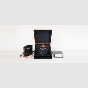
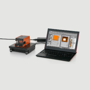
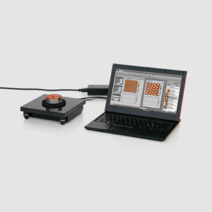
Full motorization

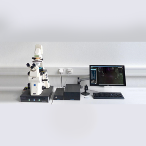
The DriveAFM is the first fully motorized AFM system that can be integrated with an inverted optical microscope. The adjustment of the two light sources for the beam deflection detection system and the CleanDrive photothermal excitation, as well as the photodetector, are fully motorized and can be controlled from the software. The tip approach to the sample is also motorized. The full motorization not only contributes to the ease of use but also allows new possibilities to fully automate the system.

DriveAFM imaging modes

This overview shows which modes the instrument is capable of. Some modes may require additional components or software options. For details, please view the brochure or contact us directly.


Standard imaging modes
Static Force Mode
Lateral Force Mode
Dynamic Force Mode (Tapping Mode)
Phase Imaging Mode

Magnetic properties

Magnetic Force Microscopy

Electrical properties

Conductive AFM (C-AFM)
Piezoelectric Force Microscopy (PFM)
Electrostatic Force Microscopy (EFM)
Kelvin Probe Force Microscopy (KPFM)


Mechanical properties

Force Spectroscopy
Force Modulation
Stiffness and Modulus
Adhesion
Unfolding and Stretching
Force Mapping
Lithography and Nanomanipulation
Electrochemical AFM (EC-AFM)
System specifications
Scan head
Scan size typ. typ. 100 µm x 100 µm x 20 µm
min. 95 µm x 95 µm x 18 µm
Read-out light source 850 nm low-coherence SLD
CleanDrive light source 785 nm laser
Photodetector bandwidth ≥8 MHz
Standard / maximum sample size 100 mm / 150 mm
Z-height noise dynamic <30 pm (RMS)
Z-height noise static <30 pm (RMS)
DC detector noise* <5 pm (RMS, 0.1 Hz – 10 kHz)
AC detector noise* <25 fm/√(Hz) above 100 kHz
Approach 10 mm motorized, parallel
(*) measured with a USC-F1.2-k7.3 cantilever
CX Controller specifications
High resolution outputs (DAC) 12x 28 bit, 1 MHz/sampling; thereof 4x user DAC (optional)
Fast outputs (DAC) 4x 16 bit, 100 MHz/sampling; thereof 1x user DAC (optional)
High resolution inputs (ADC) 12x 20 bit, 1 MHz/sampling; thereof 4x user ADC (optional)
Fast inputs (ADC) 3x 16 bit, 100 MHz/sampling; thereof 1x user ADC (optional)
Signal analyzers 2 signal analyzer function blocks that can be configured as dual channel lock-in
FPGA module and embedded processor System-on-chip module with low-latency FPGA signal processing at 100MHz and dual-core ARM processor, 2GB RAM, 1.5GHz clock
Scan control 28-bit X/Y/Z-DAC
Detector inputs Deflection/lateral signals each 20 bit
Digital sync, Spike-Guard 2-bit line/frame sync out 5 V/TTL galvanically isolated, Spike-Guard input
Clock sync 10MHz/3V clock input to synchronize data acquisition and processing
Communication to PC Gigabit Ethernet, galvanically isolated


Vous aimerez peut-être aussi…

CoreAFM-Nanosurf
Nanosurf – CoreAFM

La meilleure recherche AFM

  • Recherchez l'AFM à un prix compétitif
  • Système intégré avec un faible encombrement
  • 33 modes et fonctions
  • Manipulation facile
Le CoreAFM est le résultat de la combinaison intelligente des composants de base de l'AFM pour atteindre une polyvalence et une convivialité maximales. En raison de cette approche de conception fondamentale, le CoreAFM est équipé pour réaliser l'AFM au mieux. La fusion d'un scanner moderne guidé par flexion, d'une platine d'échantillonnage XYZ, d'une caméra, d'une table d'isolation antivibratoire active et d'un blindage du flux d'air dans une seule unité tout-en-un donne un système AFM complet avec une empreinte compacte inégalée. Le système est livré avec un contrôleur 24 bits entièrement numérique développé spécifiquement pour la tête de balayage CoreAFM. Toutes les fonctions essentielles de l'AFM moderne font partie intégrante du système CoreAFM ; tout ce que vous avez à faire pour mettre le CoreAFM en service est de connecter le contrôleur et de brancher l'alimentation et l'USB.
Lire la suite
Nanosurf - NaioAFM — AFM for small samples
Nanosurf – NaioAFM — AFM for small samples

All-in-one atomic force microscope for small samples and nanoeducation

The NaioAFM is the ideal atomic force microscope for nanoeducation and small sample measurements. This all-in-one AFM system provides solid performance and easy handling, with a price tag and footprint that fit anyone and any place.
  • Ready-to-use, table-top AFM.
  • Controller, scan head, airflow shielding, vibration isolation, camera, and sample positioning integrated into a single device.
  • All standard AFM operating modes available.
  • Integrated controller, XY-table, airflow shielding, and vibration isolation.
  • High resolution top view camera and side view sample observation built in.
  • No system setup needed: just plug into your PC and start the software.
  • Simple cantilever exchange: no adjustment required.
  • User-friendly software wizards quickly prepare measurement parameters.
  • Very competitive pricing.
[media-downloader media_id="836" texts="Click here for details"]   [embed]https://www.youtube.com/watch?v=mCUwm5TLHsw[/embed]
Lire la suite
Nanosurf - NaioSTM — STM for nanoeducation
Nanosurf – NaioSTM — STM for nanoeducation
  • Atomic resolution in minutes.
  • Extremely simple handling and reliable operation.
  • Controller and scan head integrated into a single device: just connect USB and power.
An Easy Entry into the World of Atoms The first scanning tunneling microscope (STM) was developed in 1981 by Binnig and Rohrer at the IBM Research Laboratory in Rüschlikon, Switzerland, for the first time making atoms directly visible to a small group of specialists. In 1997, Nanosurf went one step further and brought atoms to the classroom! Today, well over a thousand Nanosurf STMs play a crucial role in nanotechnology education around the globe: • Teachers appreciate the ease of use of Nanosurf STMs, allowing them to offer quick and hassle-free classroom demonstrations to their students. • Students are motivated by the rapid successes achieved when using the STMs themselves during hands-on training. • Anyone can safely handle a Nanosurf STM, since STM tips are simply cut from Pt/Ir wire without requiring etching in hazardous substances. The NaioSTM is the successor to the well-known Easyscan 2 STM and brings together scan head and controller in a single instrument for even greater ease of installation, usability, and transportability. The whole setup is very resistant to vibrations and can be used in standard classroom situations. [media-downloader media_id="837" texts="Cliquez ici pour télécharger plus de détails"]   [embed]https://www.youtube.com/watch?v=dZeZn3Pt2Ss[/embed]
Lire la suite
Nanosurf - Flex-Axiom — AFM for materials research
Nanosurf – Flex-Axiom — AFM for materials research

Versatility, performance and seamless application extensions

  • Easy to use and high performance
  • High resolution and low-noise design
  • Modes and accessories to handle all your research needs
  • Extremely modular to keep you productive
  • Upgradable for life science and operation on large stages

Applications

  • Imaging of samples in air and liquid
  • Advanced mechanical, electrical and magnetical modes
  • Electrochemical AFM
  • Environmental control
  • Scanning thermal microscopy and nano thermal analysis (SThM and Nano-TA)
  • Advanced force spectroscopy

Lateral force microscopy on polystyrene-polybutadiene blend

The lateral force imaging mode was used to image local friction differences between the two polymers

Polystyrene and polybutadiene are shown to have quite different surface properties
lateral-force-1 Overlay of difference between lateral deflection in forward and backward directions on top of topography. The difference is dominated by friction forces. Polystyrene (green areas) shows a higher friction than polybutadiene.System: FlexAFM with ES2 controller Scan size: 9 µm Cantilever: PPP-CONTSCROverlay of friction on topography
lateral-force-2 Overlay of average of lateral deflection in forward and backward directions on top of topography. The average is dominated by slope variations in the sample. The steepest slope (in red) is observed close to a large inset of polystyrene. The average shows no large color difference between the polymers in flatter areas.Overlay of slope on topography
lateral-force-3 Line section of lateral deflection difference and average of forward and backward scan directions. Line section of friction and slope signals

Electrochemical AFM

This report demonstrates the capability of the FlexAFM in studies of charged solid-liquid interfaces

In order to carry out electrochemical(in-situ)AFM experiments, a conductive sample was mounted in an electrochemical liquid cell and connected to a lab-build bipotentiostat. We employed Clavilier-type Au(111) single crystal bead crystal electrodes with facets of micrometer-wide terraces. As examples, we studied the lifting of the Au(111)-(pxV3)reconstruction, surface oxidation as well as the growth and dissolution of copper clusters in sulfuric acid solution. electrochemical-afm

3D Copper cluster grown on Au(111)

KPFM using the Nanosurf FlexAFM

Kelvin probe force microscopy imaging

Kelvin Probe Force Microscopy (KPFM) is an extension of AFM. The technique was first published in 1991 by Nonnenmacher and coworkers. Using KPFM, images can be recorded that contain information on the local work function or local contact potential difference between tip and sample. Although all Nanite systems with an SPM S200 controller and all current Easyscan 2 AFM systems are in principle capable of performing KPFM, the FlexAFM has demonstrated best KPFM performance and is therefore the instrument of choice for this type of measurement. For a detailed description of requirements and procedures, please contact Nanosurf support.
kpmg Topography. Scan Range: 10 µm x 10 µm. Z-range: 9 nm.
kpmg-2 KPFM Signal. Scan range: 10 µm x 10 µm. Local charges that were placed on an insulating (oxide) surface layer in a "Swiss cross" pattern. Image courtesy: Marcin Kisiel, Thilo Glatzel and students of the Nanocurriculum of the University of Basel.KPFM signal (top) and simultaneously recorded Topography data (bottom) are shown.
[media-downloader media_id="834" texts="Click here for details"]
Lire la suite
Nanosurf - FluidFM — Nano and cell manipulation
Nanosurf – FluidFM — Nano and cell manipulation

L'outil microfluidique pour

la nanomanipulation et la biologie unicellulaire

La microscopie à sonde FluidFM (FPM) combine la sensibilité à la force et la précision de position d'un AFM avec la technologie FluidFM de Cytosurge pour permettre toute une gamme d'applications passionnantes en biologie unicellulaire et en nanosciences. Nanosurf possède la plus longue expérience dans la fourniture de systèmes AFM avec le module complémentaire FluidFM ® , en tant que partenaire de coopération initial de Cytosurge pour cette technologie innovante - le système FlexAFM avec FluidFM ® ​​a été lancé en 2013. Les modules complémentaires FluidFM sont disponibles pour les plates-formes FlexAFM et CoreAFM et une solution FPM intégrée unique sur le FlexAFM. [media-downloader media_id="835" texts="Click here for details"]
Lire la suite